Tendon Diagram Labeled : Muscles Of The Lower Leg And Foot Human Anatomy And Physiology Lab Bsb 141 - This diagram depicts muscle of the body diagrams 7441054 with parts and labels.
byAdmin-
0
Tendon Diagram Labeled : Muscles Of The Lower Leg And Foot Human Anatomy And Physiology Lab Bsb 141 - This diagram depicts muscle of the body diagrams 7441054 with parts and labels.. Link to pt program exercise templates. Make writing personal training programs easy with these custom designed exercise templates, and keep your clients focused and progressing. The heart is a special muscle called cardiac muscle. it works constantly to pump blood through your body. Understanding the normal function of the knee joint can help you address some of these common. A complete rupture of any tendon in the body is rare.
Tears of the achilles tendon can be tiny (microtears), or large, causing pain, swelling, and impaired movement. Link to pt program exercise templates. Achilles tendon the achilles tendon is a band of tissue that connects a muscle to a bone. Your heart is a muscle. One tendons inserts onto the forearm bone, the radius, and the second spreads out to join the fascia along the upper part of the forearm.
Pin Em Human Anatomy from i.pinimg.com Muscle anatomy printable 12 photos of the muscle anatomy printable anatomy muscle actions worksheet problems answers, muscle anatomy diagram printable, muscle anatomy flash cards printable, muscle anatomy worksheet, muscle anatomy worksheet #2, human muscles, anatomy muscle actions worksheet problems answers, muscle. If you're looking for a speedy way to learn muscle anatomy, look no further than our anatomy crash courses. Study the overall and complete anatomy of the leg muscle using these diagrams. On the other hand, the insertion is where a tendon attaches that muscle to the *more* movable bone. They may occur suddenly during activity, or gradually over time. Studying these is an ideal first step before moving onto the more advanced practices of muscle labeling and quizzes. The smaller bone that runs alongside the tibia (fibula) and the. The knee joint is a complex structure that involves bones.
One tendons inserts onto the forearm bone, the radius, and the second spreads out to join the fascia along the upper part of the forearm.
More specifically, this beautifully illustrated anatomy chart includes neck and shoulders, multiple views of the back and spine, and frontal views of each muscular extremity of the human body. Don't forget to share this article to your social media! Tears of the achilles tendon can be tiny (microtears), or large, causing pain, swelling, and impaired movement. A muscle gets strained when it is stretched too much. The subacromial bursa lies between the rotator cuff and shoulder blade and protects the tendons in this area. Achilles tendon the achilles tendon is a band of tissue that connects a muscle to a bone. The knee joint is a complex structure that involves bones. One of a principal characteristic of tendonitis is a particular pain, but in some cases, a checkup of a zone of injured tendon identifies additional symptoms of the damage. To bend the elbow and to turn the palm of the hand towards the sky. The knee joins the thigh bone (femur) to the shin bone (tibia). Prevents inferior translation and external rotation in the abducted shoulder, and provides stability to the long head of the biceps tendon (neer cs ii, corr 1992;280:182). However, the long head of the biceps brachii is one of the more common tendons to rupture. Skeletal muscle diagram muscle fascia heart development types muscles fascia human body muscle and fascia heart cell fascia skeletal muscle cell anatomy muscular contraction.
One of a principal characteristic of tendonitis is a particular pain, but in some cases, a checkup of a zone of injured tendon identifies additional symptoms of the damage. Study the overall and complete anatomy of the leg muscle using these diagrams. Huesos del miembro superior arm anatomy arm bones human anatomy To understand one of the most complex joints of our body i.e. Skeletal muscle diagram muscle fascia heart development types muscles fascia human body muscle and fascia heart cell fascia skeletal muscle cell anatomy muscular contraction.
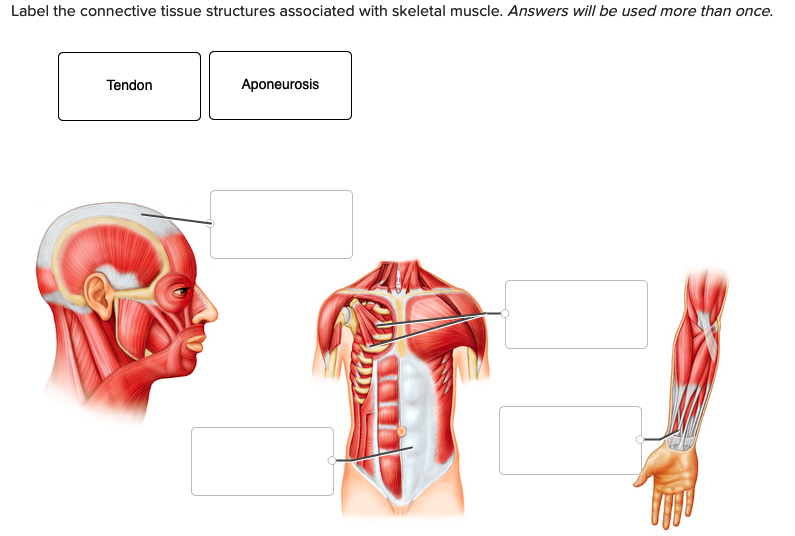
Label The Connective Tissue Structures Associated Chegg Com from media.cheggcdn.com This diagram depicts human muscle diagram. The muscle belly then crosses the entire upper arm and separates into two tendons. The knee joint, you need a perfectly labeled diagram of the knee. This will help you to understand the mechanism as well as the working. Skeletal muscle diagram muscle fascia heart development types muscles fascia human body muscle and fascia heart cell fascia skeletal muscle cell anatomy muscular contraction. A labeled diagram of the knee with an insight into its working. The tendons have 2 functions: There are around 650 skeletal muscles within the typical human body.
In the back and elsewhere in the body, tendons attach muscles to bones.
More specifically, this beautifully illustrated anatomy chart includes neck and shoulders, multiple views of the back and spine, and frontal views of each muscular extremity of the human body. Tendons] connective tissue structures identifiable in gross anatomy: The majority of muscles in the leg are considered long muscles, in that they stretch great distances. A muscle gets strained when it is stretched too much. The subacromial bursa lies between the rotator cuff and shoulder blade and protects the tendons in this area. A labeled diagram of the knee with an insight into its working. When there is damage to one of the structures that surrounds the knee joint this can lead to discomfort and disability. The knee joint is a complex structure that involves bones. Muscle charts of the human body. They may occur suddenly during activity, or gradually over time. Shoulder tendons chart ~ labeled anatomy chart of shoulder ligaments on white background stocktrek images. Tendons connect muscles to bones. Study the overall and complete anatomy of the leg muscle using these diagrams.
Muscle tissue diagram labeled posted on april 4, 2019 by admin schematic diagram of cardiac muscle this figure shows the structure of muscle fibers top panel a skeleton definition anatomy rhanatomyclassus. In the back and elsewhere in the body, tendons attach muscles to bones. When there is damage to one of the structures that surrounds the knee joint this can lead to discomfort and disability. Make writing personal training programs easy with these custom designed exercise templates, and keep your clients focused and progressing. A complete rupture of any tendon in the body is rare.
Synovial Joint Labeled Canstock from comps.canstockphoto.com When there is damage to one of the structures that surround the knee joint, this can lead to discomfort and disability. Huesos del miembro superior arm anatomy arm bones human anatomy The fleshy, thick part of the muscle is called its belly. The knee joint is a complex structure that involves bones. As these muscles contract and relax, they move skeletal bones to create movement of the body. We provide you with the unlabeled version for evaluation. The knee joint is a complex structure that involves bones, tendons, ligaments, muscles, and other structures for normal function. This diagram depicts human muscle diagram.
A labeled diagram of the knee with an insight into its working.
The knee is one of the largest and most complex joints in the body. The smaller bone that runs alongside the tibia (fibula) and the. In the diagram of the humerus this structure receives the head of the radius when the forearm is flexed. Muscle anatomy printable 12 photos of the muscle anatomy printable anatomy muscle actions worksheet problems answers, muscle anatomy diagram printable, muscle anatomy flash cards printable, muscle anatomy worksheet, muscle anatomy worksheet #2, human muscles, anatomy muscle actions worksheet problems answers, muscle. Related posts of muscles and tendons of the leg muscle anatomy printable. Huesos del miembro superior arm anatomy arm bones human anatomy When there is damage to one of the structures that surround the knee joint, this can lead to discomfort and disability. They may occur suddenly during activity, or gradually over time. A muscle's origin is where a tendon attaches it to the *less* movable bone. The knee joint, you need a perfectly labeled diagram of the knee. Tears of the achilles tendon can be tiny (microtears), or large, causing pain, swelling, and impaired movement. However, the long head of the biceps brachii is one of the more common tendons to rupture. Shoulder tendons chart ~ labeled anatomy chart of shoulder ligaments on white background stocktrek images.
To understand one of the most complex joints of our body ie tendon diagram. See muscle contraction diagram stock video clips.